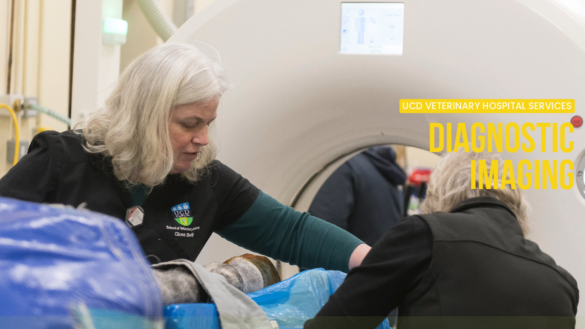
Computed Tomography (CT) is a very versatile diagnostic imaging technique that uses X-radiation to create tomographic images of the body; the result is a 3D image of any organ/structure of the body from a carpus joint to a porto-systemic shunt. The UCDVH has the only CT scanner in the Republic of Ireland for veterinary use, a SOMATOM SCOPE 16 slice moving gantry capable of doing standing equine patients as well as small animals.
The clinical indications for performing CT scans are almost endless, and range from nasal disease to abdominal masses and elbow dysplasia in small animals to dental disease and sinusitis in horses. The procedure times are usually very short and in many cases sedation is sufficient.
CT scans are mostly performed on inpatients; however, we also provide a service for direct external referrals from veterinary surgeons. This is ONLY dedicated to elective scans in dogs that are clinically stable and DO NOT need to be anaesthetized for the scan (e.g. investigation of elbow dysplasia or shoulder osteochondrosis). The cost of this service varies with the area of interest and can be booked by calling the Diagnostic Imaging unit directly on (01) 716 6098.

Almost 5,000 Small Animal and 1,000 Large Animal cases go through the UCDVH Diagnostic Imaging (DI) unit each year. The unit provides a full range of Diagnostic Imaging services for all internal patients admitted to the Hospital, but also a variety of services for external cases referred to the UCDVH by veterinary practitioners or booked in directly by clients. All of our equipment is state of the art, veterinary dedicated and operated by our expert team.
Both inpatients and direct referral cases can avail of a cardiology service that includes a complete visit and electrocardiogram performed by one of our specialist imagers as well as our visiting cardiologist Dr Rachel Blake. Obtaining images in B-mode, M-mode the radiologist can assess the size of the cardiac chambers, presence of cardiac defects, cardiac contractility and, using Doppler technology, the velocity of blood flows and regurgitations at the valves etc. The exam is performed in a quiet, dark room, and the animal is not sedated.
This service can be booked through the UCDVH Reception at (01) 716 6000.
The specialist radiologists in our unit provide a film reading service to veterinary practitioners in need of a radiological opinion on their cases.
Radiographs can be sent in by email to (opens in a new window)vetdiagnosticimaging@ucd.ie
The price for oral report is €30 and the response time is usually within 24 hours.
Fluoroscopy allows us to assess internal structures of the body, similar to a radiograph, but whilst moving. Because the images are immediately available, fluoroscopy can be used in surgical theatres during orthopaedic surgeries to assess the placement of implants, such as screws and plates. The UCDVH has a C-arm Siremobil 4K and Ziehm Vision Vascular machines that can be easily moved to various locations within the Hospital as required.
Our MRI unit is a high field 1.5 tesla GE SIGNA ARTIST with an array of specialised radiofrequency coils. MRI is the technique of choice for investigating neurological (seizures, blindness, ataxia etc) and musculotendinous disorders. The UCDVH has a state-of-the-art MRI facility on-site, which provides top quality images for investigation of brain, spinal cord, nerves, musculotendinous structures and abdominal organs.
MRI scans are mostly performed on inpatients and always under general anaesthesia; in exceptional cases, we accept direct referrals from veterinary practices. The cost of this service varies with the area of interest; enquiries and bookings can be made by calling the Diagnostic Imaging unit directly on (01) 716 6098.
Our state of the art equipment includes:
- Small animal Direct Radiography system: Siemens Axiom Aristos FX Plus DR with moving tube and detector
- Large animal system: Siemens Vertix Vet that can be used with both Direct Radiography and Computer Radiography
- A portable machine
- Three mobile machines
The recent acquisition of two portable DR plates has added great versatility to both small and large animal radiography. In equine radiography, they have reduced greatly the time required to complete a radiographic study and therefore the amount of sedation needed for the horse; furthermore they can also be used to perform intra-operative studies or on cases where a patient cannot be moved from ICU or isolation for welfare/safety reasons.
From abdominal organs, to thyroids, hearts and tendons, ultrasound can provide useful information across a range of cases. In the UCDVH Diagnostic Imaging unit, there are two ultrasound suites; one to perform abdominal studies with a dedicated GE Logic 9 and 4 transducers 4-10, 8-15, 3-5 MHz; and one to perform echocardiographic studies with a dedicated GE Vivid.
We also have two smaller, portable US machines: a BCF Micromax with two transducers (6-13Mhz linear phased array and 1-5 sector phased array) and a BCF Micromax Turbo with two transducers (6-13Mhz linear phased array and 1-5 sector phased array). These machines are mostly used for equine abdominal and musculoskeletal scanning, but also when we are called out to cases in Dublin Zoo.
Once a month we host a cardiology day with Dr Rachel Blake a specialist cardiologist, who performs cardiac scans for certifications for breeds such as Newfoundland, Bernese Mountain dogs, Dobermanns etc. Dr Blake also reviews specialist cases from the UCDVH.