On this page:
- Andor spinning disk confocal
- Critical Point Dryer
- CytoViva
- Epi-fluorescent microscope for dishes
- Epi-fluorescent microscope for dishes
- Epi-fluorescent microscope for slides
- FLIM microscope
- Glow Discharger/sputter coater
- Harmony HCS/HCA image analysis workstation
- High-pressure freezer
- Leica DMI6000B epi-fluorescent microscope
- Leica UC6 Ultramicrotome
- Leica UC7 Ultramicrotome
- Low-magnification microscope
- Nanotom M X-ray CT scanner
- Nicolet iN10 MX
- Olympus FV1000 confocal microscope
- Olympus FV3000 confocal microscope
- Olympus ScanR epi-fluorescent HCS microscope
- Opera Phenix confocal HCS microscope
- OPTIR-AFM-IR
- Raman
- Spero
- Spinning disk confocal microscope
- Sputter coater
- Tecnai 12 TEM
- Tecnai 20 TEM
- Transmitted light microscope
- VG Studio MAX
- Vis-SWIR
- Vtomex M 240 X-ray CT scanner
- Zeiss LSM 800 Airy
- Zeiss Sigma 300 SEM
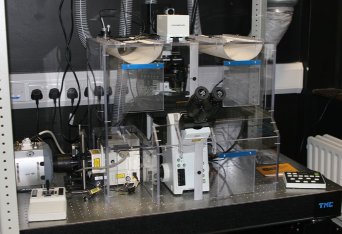
Andor spinning disk confocal
Advanced inverted microscope for live cell imaging. Real confocal images up to 30 frames per second at full frame resolution. Four lasers (445nm, 488nm, 514nm, 561nm), minimal photobleaching and phototoxicity. Ultimate sensitivity of Andor iXonEM EMCCD camera. Rapid optical sectioning for 4D imaging. Powerful visualisation, image analysis and tracking tools using IQ software.
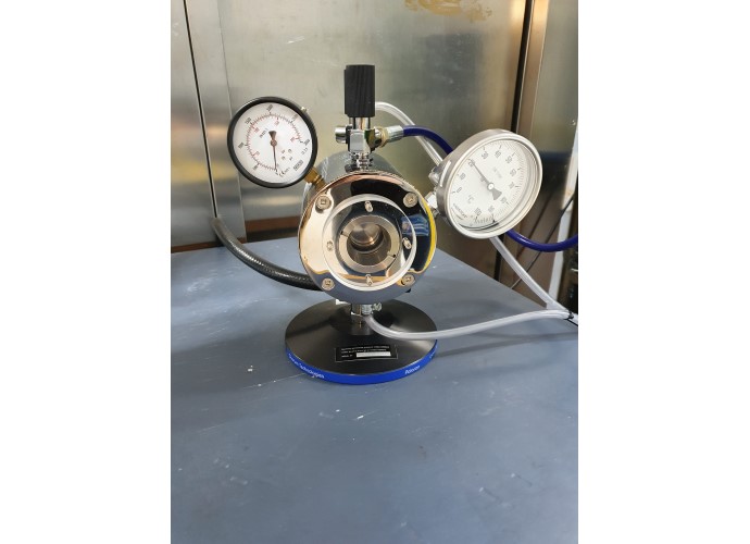
Critical Point Dryer
The E3100 large chamber critical point dryer has been an industry standard for over 35 years and is used in numerous scanning electron microscopy (SEM) laboratories around the world. Primarily used for critical drying of biological and geological specimens, the E3100 can also used for the controlled drying MEMs, aerogels and hydrogels.
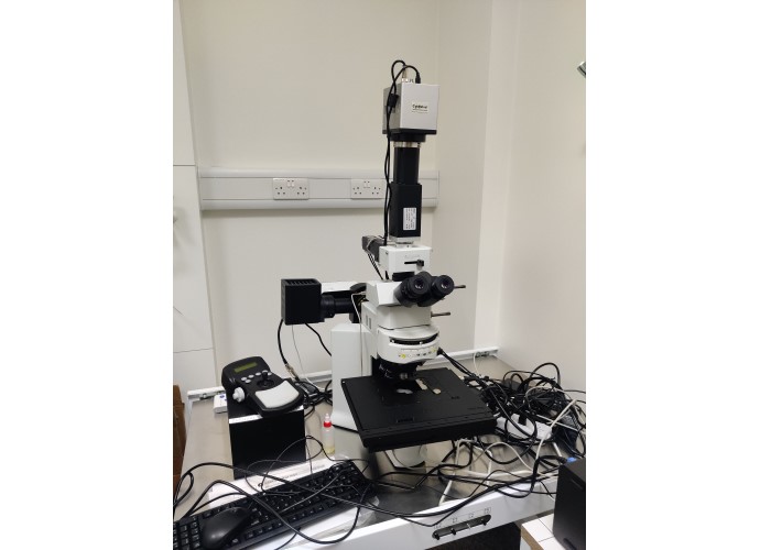
CytoViva
CytoViva provides hyperspectral imaging solutions from the visible to near infrared range, which are specifically designed to work with all modalities of optical microscopy. This includes CytoViva’s patented enhanced darkfield microscope optics, fluorescence and standard brightfield microscopy.
A hyperspectral microscope is unique in its ability to capture optical spectral data in every pixel of the microscopic sample image. The hyperspectral microscope enables comparative spectral review, spectral mapping and other analysis of different elements within a sample. These sample materials can include fluorescent, plasmonic or other light scattering materials as well as live cell and tissue samples.
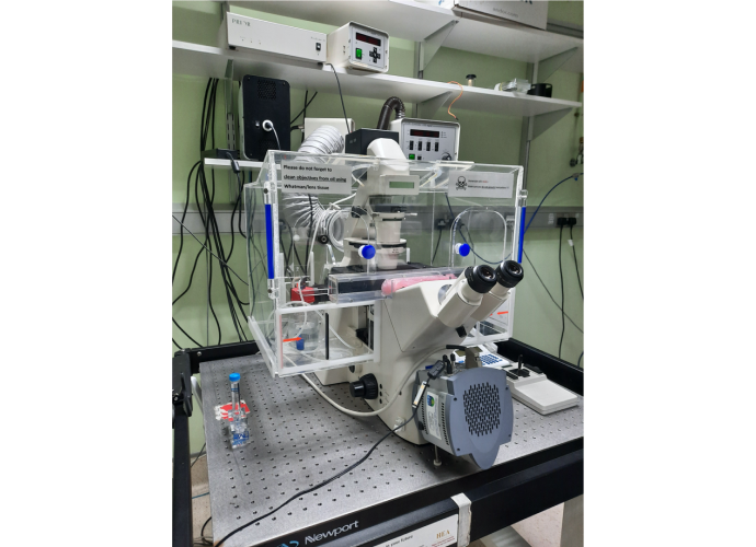
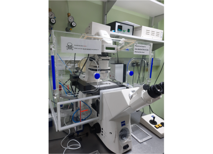
Epi-fluorescent microscope for dishes
An inverted Zeiss Axiovert 200 microscope for imaging of live and fixed cells grown in culture dishes. For multiplex epi-fluorescent microscopy in the whole visible spectrum and near infra-red. Optional: transmission light microscopy. Optional: multi-positional time-lapse detection. Marina
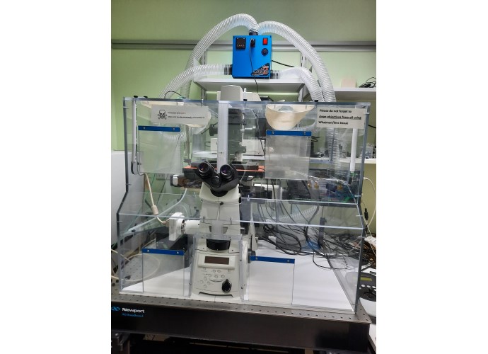
Epi-fluorescent microscope for dishes
An inverted Zeiss Axiovert 200 microscope for imaging of live and fixed cells grown in culture dishes. For epi-fluorescent microscopy in the whole visible spectrum and near infra-red. Optional: transmission light microscopy. Optional: multi-positional time-lapse detection. Suzi
FLIM microscope
Microscope dedicated to fluorescent live imaging (FLIM), frequency domain. Integrated by Lambert. Optional: high-power high-resolution transmission light microscopy for live cells
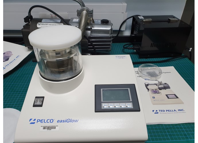
Glow Discharger/sputter coater
The PELCO easiGlow™ Glow Discharge Cleaning System is a compact, quick and easy to use standalone system. It is primarily designed for cleaning TEM grids and hydrophilization of TEM carbon support films, which have the tendency to be hydrophobic. A glow discharge treatment with air will make a carbon film surface negatively charged (hydrophilic) which allows aqueous solutions to spread easily.
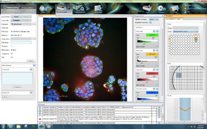
Harmony HCS/HCA image analysis workstation
The Perkin Elmer Harmony image analysis software offers state-of-the-art single cell analysis and quantification possibilities from high numbers of samples across multi-well plates. Analysis 'pipelines' are completely user-defined, consisting of several stages. Typically each cell in every well is identified (segmented), and then depending on the fluorophores present in the sample, a range of per cell features are extracted and quantified.
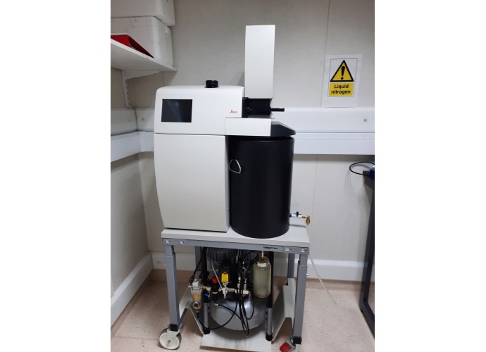
High-pressure freezer
High-pressure freezer is a method of fixation of biological objects (cell culture, pieces of tissue, small animals like C.elegans, etc.) for electron- and correlative microscopy.
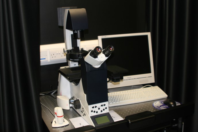
Leica DMI6000B epi-fluorescent microscope
Inverted wide-field microscope for fluorescence cell imaging. Equipped with six filters and six objectives, a scanning stage, and a high-resolution CCD camera for easy and rapid cell imaging.
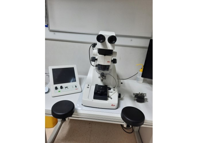
Leica UC6 Ultramicrotome
The Ultramicrotome Leica EM UC6 provides easy preparation of semi- and ultrathin sections as well as perfect, smooth surfaces of biological and industrial samples for TEM, SEM, AFM and LM examination.
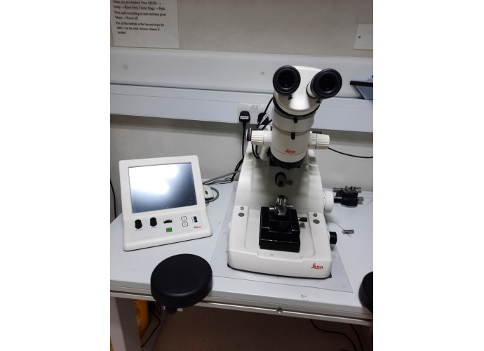
Leica UC7 Ultramicrotome
The Ultramicrotome Leica EM UC7 provides easy preparation of semi- and ultrathin sections as well as perfect, smooth surfaces of biological and industrial samples for TEM, SEM, AFM and LM examination.
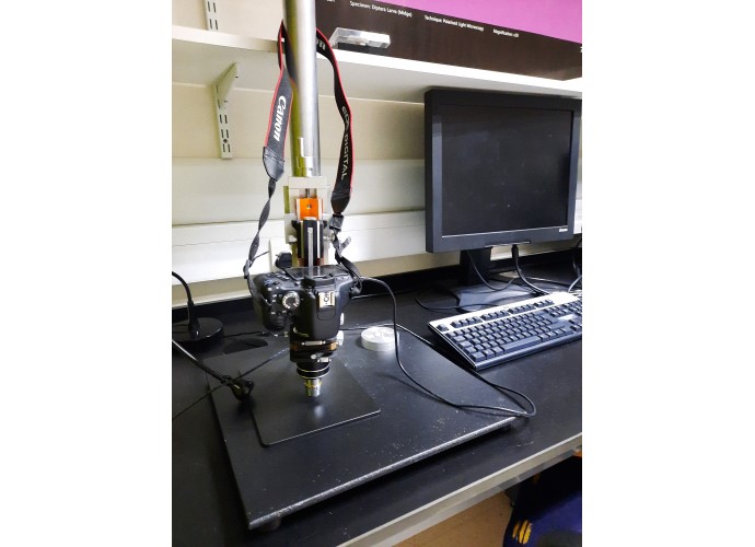
Low-magnification microscope
A basic low-magnification microscope with high-resolution camera
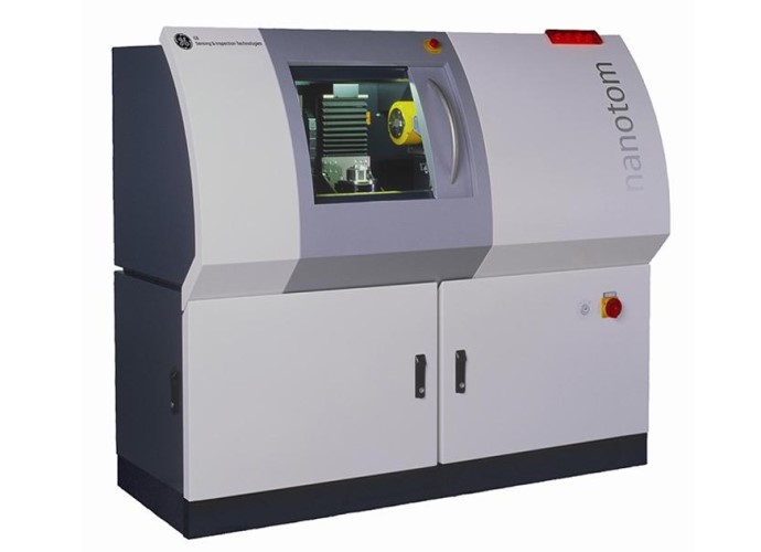
Nanotom M X-ray CT scanner
Micro X-ray CT scanner. 180 kV. Can scan <1 um. We can scan any sized sample from a poppy seed to a cola can. Scans take approx 9 mins. Access charge model in place.
Contact: (opens in a new window)Saoirse Tracy
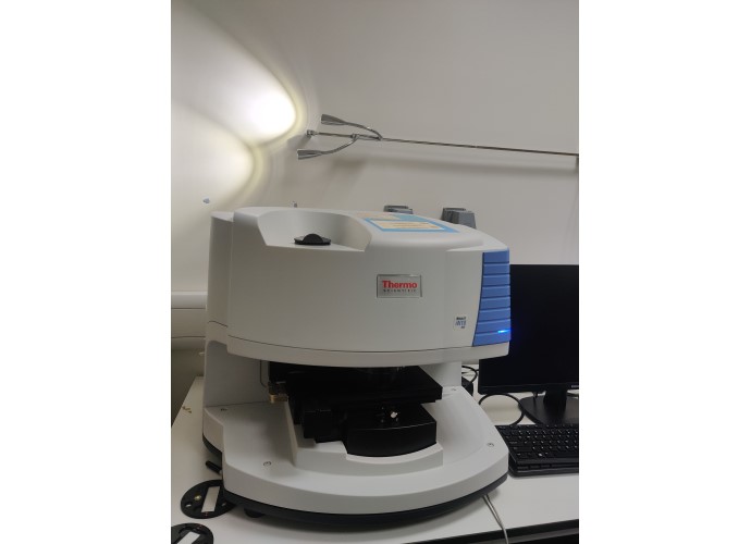
Nicolet iN10 MX
The Nicolet iN10 MX Infrared Imaging Microscope provides the power required to rapidly acquire and analyze chemical images to enhance your understanding of the chemical distribution of materials in heterogeneous samples.
- Microspectroscopy
- Material identification
- Packaging and laminate
- Coating
- API mixture distribution mapping
- Contaminant identification
- Failure analysis
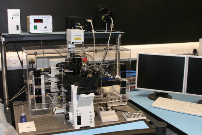
Olympus FV1000 confocal microscope
Advanced inverted IX81 microscope for confocal imaging. Point scanning confocal images up to 4096 x 4096 resolution. Six lasers (405nm, 445nm, 488nm, 514nm, 559nm, 640nm) and twin scanners allowing simultaneous bleaching and acquisition. Detection on five photomultipliers, two equipped with spectral detection. Climate control system (temperature, humidity, CO2) for long-term live imaging. Powerful visualisation, image analysis and tracking tools using Olympus Fluoview software. Free support available to all users.
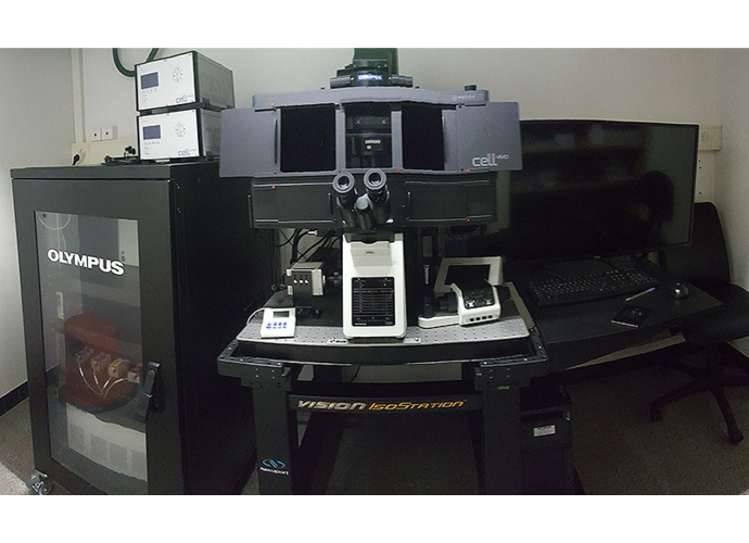
Olympus FV3000 confocal microscope
An inverted Olympus IX83 microscope, with 7 laser lines all over the visible spectrum (405, 445, 488, 515, 560, 595 and 640 nm). Best for multiplex confocal imaging of live cells and micro-organoids.
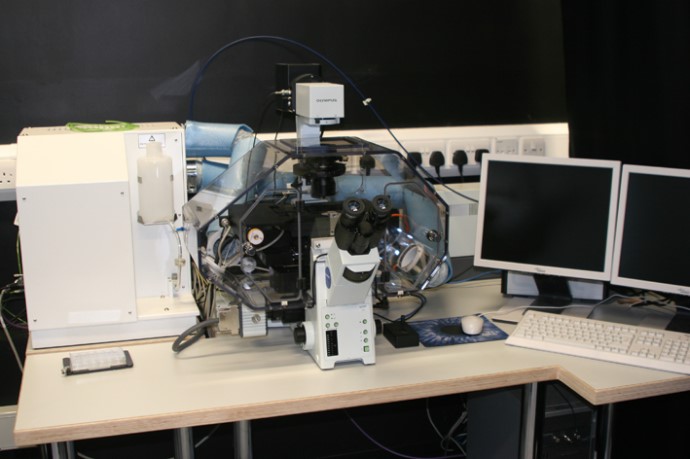
Olympus ScanR epi-fluorescent HCS microscope
Advanced fully automated inverted wide-field microscope for high content screening applications. Xenon light source for quantitative imaging, rapid excitation filter wheel, fast filter turret, detection on 1344 x 1024 resolution cooled CCD camera. Fully automated objective changing, stage function, autofocus (hardware- and software-based) and acquisition. Climate control system (temperature, humidity, CO2) for long-term live imaging.
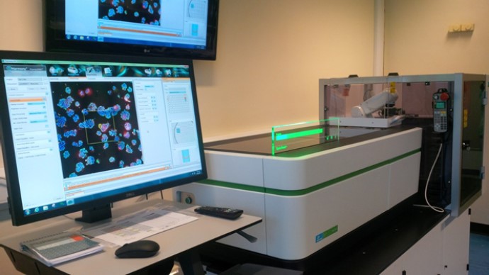
Opera Phenix confocal HCS microscope
Advanced fully automated inverted spinning disk confocal microscope for high content screening (HCS) applications. Four excitation LED laser lines (405, 488, 561 and 640 nm), six objectives (5x to 63x, of which three are air and three are water-immersion), four parallel sCMOS cameras (16 bit, 4.4 mega pixel, 2100 x 2100 resolution, 6.5 µm pixel size). Climate control system (temperature, humidity, CO2) for long-term live imaging. Robotic loading arm for automated plate loading; automation scheduling software. Room temperature 'plate hotel' with 14 positions. Integration with Liconic CO2 incubator containing 'plate hotel' with 44 positions. Seemless image transfer to Harmony and Columbus image analysis platforms.
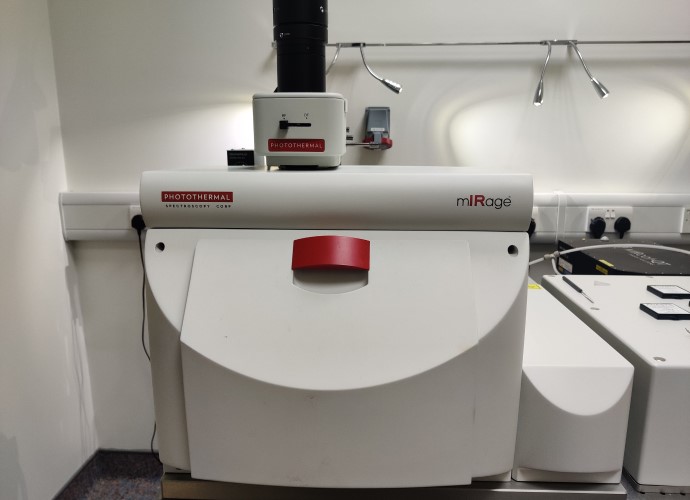
OPTIR-AFM-IR
The mIRage is sub-micron IR spectroscopy and imaging system. OPTIR is a proprietary technique that breaks the diffraction limit of infrared. It bridges the gap between conventional IR micro-spectroscopy and nanoscale IR spectroscopy.
- Sub-micron IR spectroscopy and imaging
- Non-contact – fast and easy to use
- Transmission quality IR spectra in reflection mode
- No need for thin sections
Raman
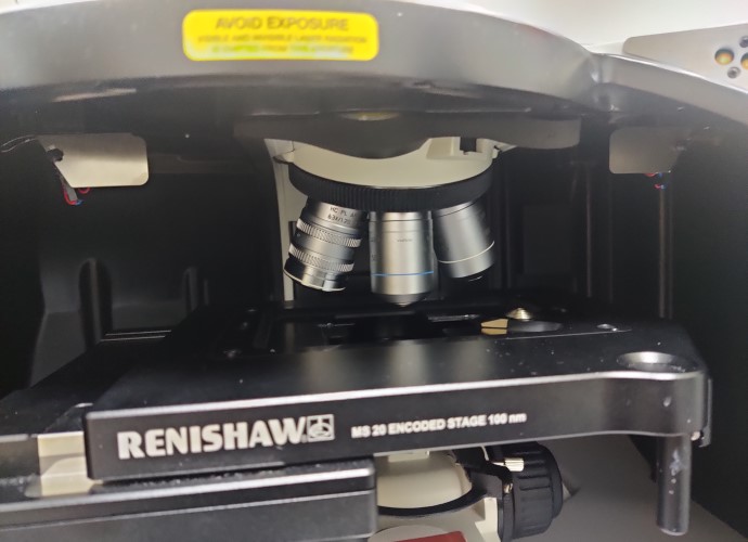
inVia Raman microscope
- high throughput and high sensitivity
- can resolve weak Raman bands (4th order silicon)
- high spectral and spatial resolutions
- 4 objectives: 10x (0.25 NA), 50x (0.75 NA), 63x (1.2 NA) and 100x (.085 NA)
- 2 lasers: 785 nm and 532 nm
Spero
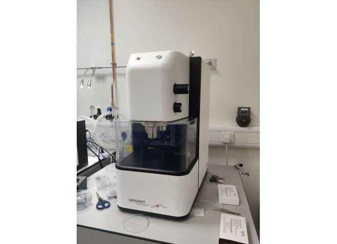
Spero Chemical Imaging Microscope. High throughput and high sensitivity, label free infrared microscopy
- Transmission, visible and reflection modes
- Diffraction limited, high-sensitivity imaging with Focal Plane Array (FPA) detector
- Multiple, high-NA, large FOV imaging optics (0.7 NA and 0.3 NA)
- Live, real-time infrared imaging
- High-throughput hyperspectral imaging enabled by ultra-high brightness QCL technology (>7 M spectral points per second)
- Large, flexible sample compartment
- Multiple configuration options including extended wavelength coverage and automated polarization control
Spinning disk confocal microscope
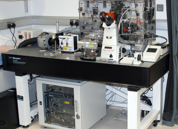
An inverted Nikon Ti microscope with 5 laser lines and Yokogawa spinning disk integrated by Andor Fusion. For confocal microscopy of live cells. Optional: transmission light microscopy. Optional: multi-positional time-lapse detection. Optional: SRFF-super-resolution. Optional: TIRF microscopy
Sputter coater
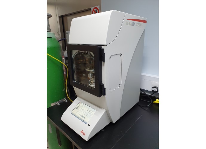
Sputter coater is essential for sample preparation for scanning electron microscopy (SEM), if secondary electron (S2) or In-Lens detectors are used.
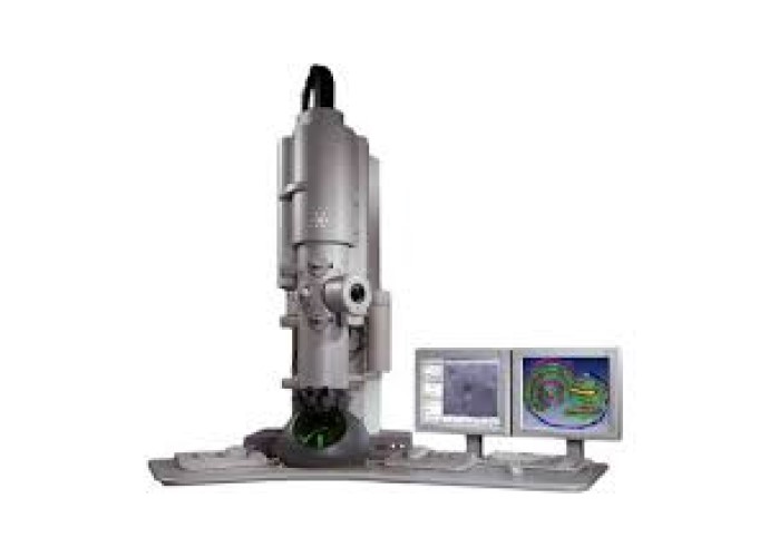
Tecnai 12 TEM
Tecnai 12 is an easy to use 20 kV to 120 kV transmission electron microscope (TEM) designed to provide high-contrast, high-resolution imaging and analysis.
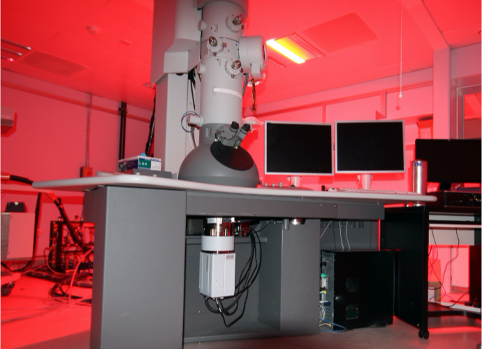
Tecnai 20 TEM
Tecnai 20 is a transmission electron microscope (TEM) with electron acceleration from 20 kV to 200 kV designed to provide high-contrast, high-resolution imaging and analysis.
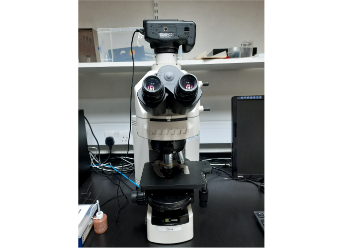
Transmitted light microscope
A dedicated transmission light microscope with a high-resolution power. Optional: differential interference contrast (DIC), dark filed microscopy, polarized microscopy. Orina
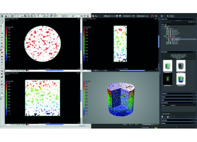
Vis-SWIR
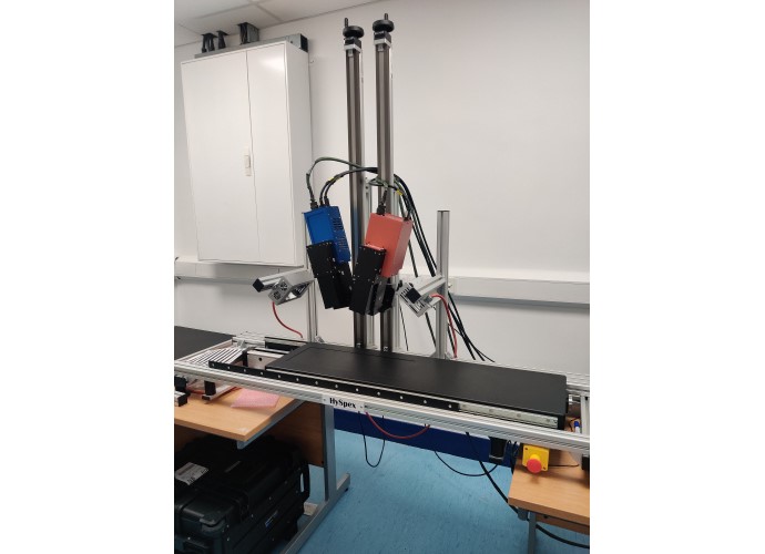
Visible - Short wave infrared (Vis-SWIR) Hyperspectral imaging enhances understanding of biological through spatially resolved measurement of their interaction with light. This system includes:
- FX10e VNIR 400-1000 nm hyperspectral camera
- Specim SWIR-CL-400-N25E SWIR 1000-2500 nm hyperspectral camera
- SWIR lens
Vtomex M 240 X-ray CT scanner
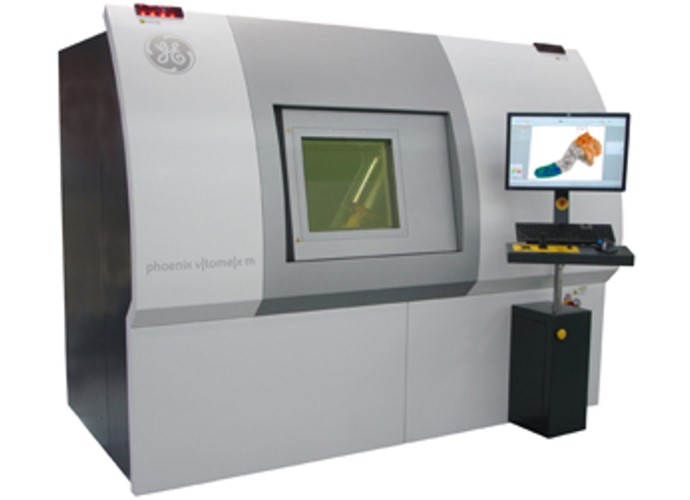
Micro X-ray CT scanner. 240 kV. Can scan <10 um. Max sample diameter 15 cm and approx 50 cm long. Scans take approx 9 mins. Access charge model in place.
Contact: (opens in a new window)Saoirse Tracy
Zeiss LSM 800 Airy
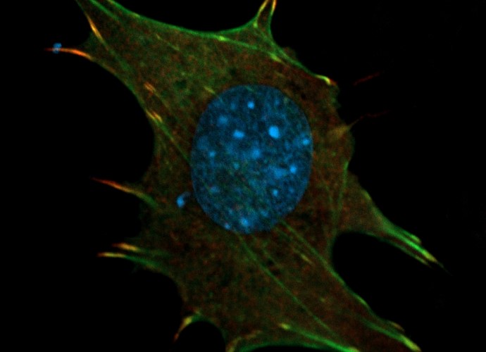
An upright confocal and Airy scan microscope with four laser lines (405, 488, 560, and 640 nm), with a motorized stage. Ideal for imaging of biological samples on slides, for scanning of larger areas and volumes, and for the correlative light-electron microscopy.
Zeiss Sigma 300 SEM
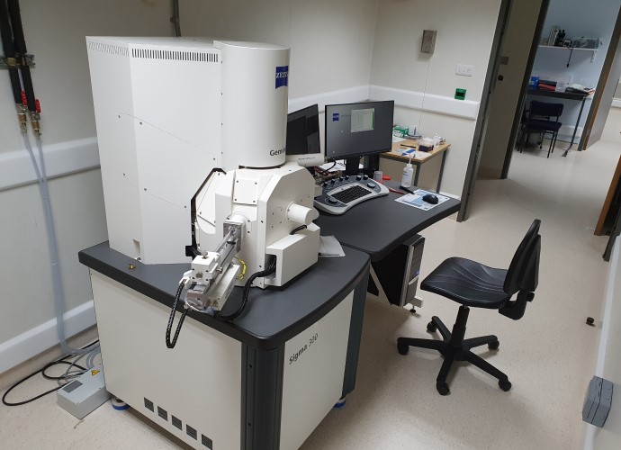
Zeiss Sigma 300 is a brand-new high-vacuum FEG-SEM electron microscope equipped with four detectors and the resolution of 1.2 nm, with the software for correlative light-electron microscopy.